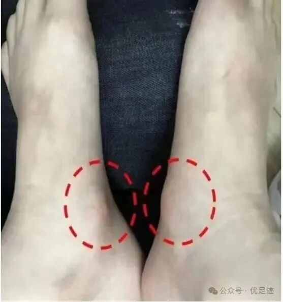
Recently, many children have come in with a painful protrusion on the inner side of the foot. Experienced assessors will first suspect accessory navicular pain. Here is some relevant information about this condition for parents.
Classification of Accessory Navicular Bone
The accessory navicular bone is divided into three types morphologically:
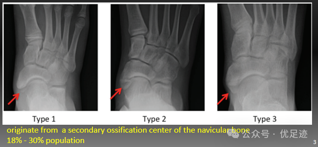
Type 1: A small round sesamoid bone embedded within the tendon, with a diameter of 2 to 3 millimeters. It causes no bony impingement or friction against the navicular bone, with a clearance of over 3 millimeters between them.
Type 2: A separate accessory navicular bone that appears triangular on X-ray. It generally measures about 8 to 10 millimeters in size and may be larger in adults. It connects to the navicular bone via fibrocartilage, with a clearance of less than 2 millimeters.
Type 3: Resembles an enlarged navicular tuberosity. The navicular bone presents a horn-like shape, forming an enlarged medial tuberosity together with the accessory navicular.
Pathogenesis of Accessory Navicular Pain
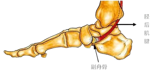
The accessory navicular is the secondary ossification center of the navicular bone. During development, this ossification center fuses with the navicular bone in most people. If fusion fails, a separate accessory navicular forms, which is connected to the navicular bone by fibrocartilage.
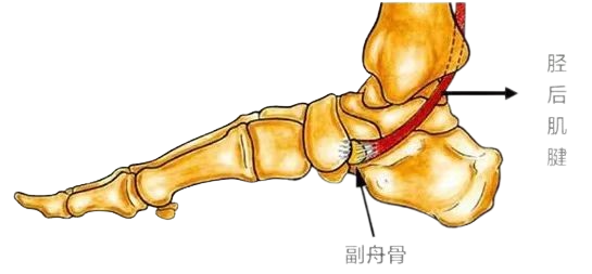
The posterior tibial tendon usually wraps around or attaches to the accessory navicular bone. The posterior tibial muscle has multiple insertion points around the navicular bone, among which the area of the accessory navicular and navicular bone bears the greatest load and serves as a major pathway for stress transmission.
As the second strongest muscle in the lower leg, the posterior tibial muscle generates tremendous compression, tension and shear force locally. It acts as the primary muscle for foot inversion, stabilizes the midfoot and hindfoot, absorbs impact on the feet, and works together with the triceps surae to propel the body forward.
During movements that require forefoot propulsion such as running, sprinting, jumping and rope skipping, the posterior tibial muscle exerts intense force.
Pain occurs when these mechanical movements cause injuries to the fibrocartilaginous junction. Common triggers include ankle sprains, trauma and friction from footwear.
Patients diagnosed with accessory navicular syndrome who suffer from pain, swelling and other symptoms after exercise or prolonged standing, with daily life severely affected, usually require surgical intervention when conservative treatments fail to relieve their conditions. Since Kidner first proposed surgical treatment for symptomatic accessory navicular bones in 1929, a variety of surgical techniques have been developed. Traditional procedures mainly include simple excision of the accessory navicular bone, the Kidner procedure and its modified versions, as well as accessory navicular-navicular fusion.
Restricting activities, elevating the affected limb, extracorporeal shock wave therapy and anti-inflammatory medication can relieve local inflammation and swelling of the foot and ankle, and ease pain around the accessory navicular bone. However, these rehabilitation measures only address fascial inflammation, swelling and adhesion caused by the accessory navicular deformity, and raise the pain threshold. They have limited effects on correcting abnormal arch structure, plantar stress points, foot stability, biomechanical alignment, as well as impaired energy storage and release during movement. Consequently, pain around the accessory navicular bone often recurs after exercise and undermines patients' quality of life.
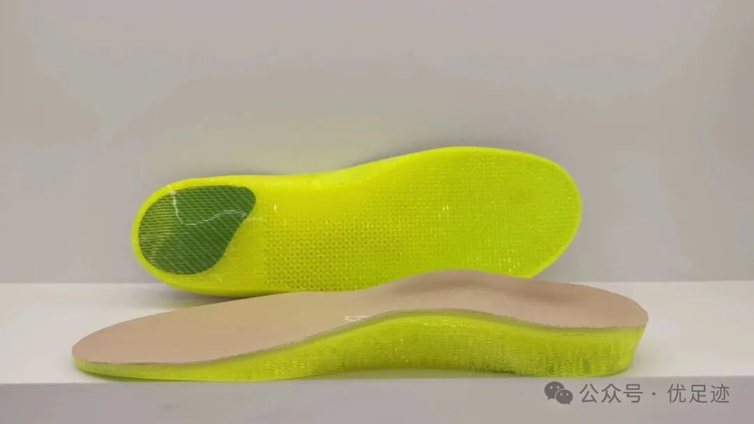
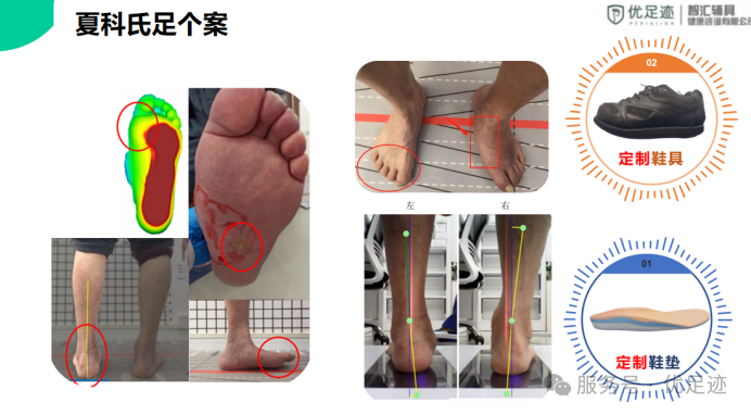
For this reason, custom orthotic insoles play a vital role. Equipped with arch supports, they provide proper arch support, redistribute plantar pressure and restore normal biomechanical alignment to alleviate pain. Additionally, they improve cutaneous sensation and proprioception, enhance balance and correct abnormal gait.